File Decode

For our reconstruction, actual tomographic data from a gamma camera was obtained. The images were of a standard phantom, which in our case, was of a cylinder filled with hot and cold rods. This data was obtained courtesy of Dr. Bud Wendt, from the M.D. Anderson Center in Houston, TX.
The data came in Interfile 3.3 format, which is a standard data format for used in Nuclear Medicine. It consists of two files: a descriptive ASCII header file, and a purely binary data file. The header gives important details about the images in the data file; for example, who the patient was, what imaging machinery was used, and the sizes and number of images. The data file consists of raster images, scanning from left to right and top to bottom. For more information, the details of the Interfile standard can be found here.
The datafile consisted of 180 128x128 pixel images, each being a projection of the phantom dummy at a different angle. The projections went completely around the dummy, as one was taken for every two degrees of rotation.
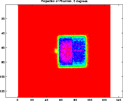
The above image is one of the projections of the phantom, this one being from directly over the top. The axis of the gamma camera runs from left to right, while the camera spins from top to bottom. Note that the camera takes a two-dimensional image at every angle; with this information, we want to reconstruct a two-dimensional slice through the dummy, perpendicular to the projection planes.
For us to calculate the tomographic slices, we needed to take a column from each projection and group them all together. The following sinogram is a collection of all the 64th columns from all of the projections; the columns are then rotated to be horizontal.
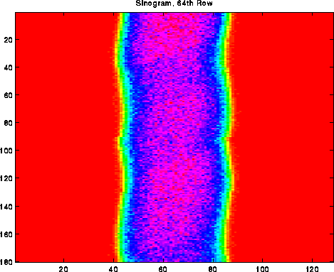
Each one of the rows is a vector from one of the projections; this vector runs perpendicular to the axis of the phantom body.
To create the sinogram, represented in MATLAB as a 128x180 element matrix, some MATLAB code was written. The function can be found here.
| On to the next page... | Back to the front page. |