 |
Experience is one thing you can't get for nothing. |
Home
Lab Resources
- Agarose Gel Electrophoresis
- Bacterial Streak Plate
- Bacterial Transformation
- DNA Ligation
- PCR
- Pipettors
- Plasmid DNA Isolation and Restriction Enzyme Digests
- Preparation of Agar Plates
Pre-lab Preparation
- Laboratory Citizenship & Performance
- BioSciences Lab Program Honor Code
- Molecular Biology Tips
- Writing Up Methodology
Additional Resources
Bacterial Transformation
Introduction of Foreign DNA into Cells
Foreign DNA can be placed in cells by several methods. If the foreign DNA is introduced into the cell in a form acceptable to the host, genes on that DNA can be expressed and the DNA can be propagated by the cells. In many cases this is done by attaching the foreign DNA to a piece of DNA that is capable of replicating within the host. For bacteria and some eukaryote species, plasmids or phages represent suitable vectors. Plasmids can carry only relatively small segments of DNA (<15 kb) but phage or cosmid vectors can carry up to 50 kb. Yeast artificial chromosomes (YAC) can be used to propagate very large foreign DNA fragments (>200 kb) in yeast. Phage or virus particles are available to transfer DNA into almost any type of cell. In other cases, foreign DNA can be introduced without any attached vector, and can sometimes integrate itself into the host chromosome where it is replicated as part of the host genome.
The overall process of changing the phenotype of a bacterium
by introducing a plasmid into it is called transformation (there
are other processes that are also called transformation but
we will not concern ourselves with these). Bacteria may be
transformed with plasmids by several techniques. The simplest
is merely incubating the plasmid with bacteria whose cell wall
has been weakened. Treating the bacteria with calcium or rubidium
makes the membrane permeable to DNA through an unknown mechanism;
these chemically treated cells are referred to as "competent" because
they are now ready to take up foreign DNA. Special strains
are also available with genetic alterations that limit the
formation of the polysaccharide layer making the transformation
of these cells 2 to 3 orders of magnitude more efficient. Organic
solvents (DMSO) and polyethylene glycol (PEG 8000) may be used
in transformation procedures; these methods may have slightly
lower efficiencies but are more rapid to perform. Less natural
methods of placing DNA into cells are also used. DNA attached
to microscopic particles can be physically "shot" into
cells (Ballistic transformation) or soluble DNA can enter by
blasting holes in the cell membrane by a high-voltage electric
discharge (electroporation). The method of choice depends on
the type of cell and the instrumentation available.
Bacterial cells can also be transformed by electroporation.
Electroporation involves exposing a suspension of cells and DNA
to a high-voltage electric discharge, which creates pores in
the cell membrane. These pores are large enough to allow DNA
entry into the cell. Physiologically, the cell membrane, which
acts as a capacitor, is unable to pass current except through
ion channels; subjecting the membrane to a high-voltage electric
field causes a temporary breakdown in membrane integrity, and
the reclosing of the holes is a natural decay process. Transformation
using this method is very efficient (several orders of magnitude
greater than traditional chemical methods).
For every transformation, one or more controls should be performed:
- Positive Control -- transform competent cells with plasmid DNA (not digested); provides measure of the efficiency of transformation and serves as a standard for comparison with other transformations
- Negative Controls
- NO DNA -- transform competent cells, without added DNA, and plate on selection media; if high background, may indicate a problem with the antibiotic in the agar plates
- Digestion efficiency -- transform competent cells with digested plasmid DNA / NO ligase treatment; if high background, may indicate that the digestion did not go to completion
- Vector recircularization -- transform competent cells with digested vector DNA that was treated with alkaline phosphatase before adding ligase (NO insert is present); if high background, may indicate that the dephosphorylation reaction was incomplete
Agarose gel purification of digested plasmid and insert DNA is one way to decrease background from inefficient digestion by restriction enzymes. Using restriction enzymes that generate noncompatible "sticky" ends decreases background from vector recircularization.
- References:
- Andreason, G.L. and G.A. Evans. (1989) Optimization of electroporation for transfection of mammalian cell lines. Anal. Biochem.180 (2): 269.
- Chung, C.T. and R.H. Miller. (1988) A rapid and convenient method for the preparation and storage of competent bacterial cells. Nucl. Acid. Res. 16: 3580.
- Cohen, S.N., A.C.Y. Chang, and L. Hsu. (1972) Nonchromosomal antibiotic resistance in bacteria: Genetic transformation of Escherichia coliby R-factor DNA. Proc. Natl. Acad. Sci., USA 69: 21102114.
- Cohen, S.N., A.C.Y. Chang, H.W. Boyer, and R.B. Helling. (1973) Construction of biologically functional bacterial plasmids in vitro. Proc. Natl. Acad. Sci. USA 70: 3240-3244.
- Knutson, J.C. and D. Yee. (1987) Electroporation: parameters affecting transfer of DNA into mammalian cells. Anal. Biochem.164 (1): 44.
- Potter, H., L. Weir, and P. Leder. (1984) Enhancer-dependent expression of human kappa immunoglobulin genes introduced into mouse pre-B lymphocytes by electroporation. Proc. Natl. Acad. Sci.81: 7161.
- Sugar, I.P. and E. Neumann. (1984) Stochastic model for electric field-induced membrane pores: Electroporation. Biophys. Chem.19 (3): 211.
- Tsong, T.Y. (1991) Electroporation of cell membranes. Biophys. J. 60: 297-306.
- Weaver, J.C. (1993) Electroporation: A general phenomenon for manipulating cells and tissues. J. Cell. Biochem. 51: 426-435.
Preparation of Competent Cells
Competent cells can be purchased from commercial vendors such as New England Biolabs (e.g., NEB 5-alpha Competent E. coli). Or competent cells can be prepared in the lab. Mix & Go E. coli Transformation Kit & Buffer Set (Zymo Research, Irvine, Ca) makes it fairly easy to make competent cells from the E. coli strains you work with in your own lab.Growth and Check of Bacterial Strains
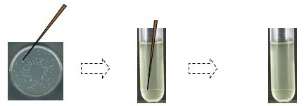
Bacteria can be propagated on liquid or solid media. The use of liquid allows large quantities of bacteria to be harvested but does not permit easy selection or determination of phenotype of single cells. The technique of "streaking" cells onto a solid media provides simple isolation of colonies arising from single cells. Colonies selected for the desired phenotype are then used to inoculate liquid broth. A single colony inoculum is preferred because bacteria can undergo many types of mutations naturally. The instability of some of the mutations, especially transposons and phages, can allow some cells to lose characteristics important to the selection scheme and may complicate the analysis. It is always wise to check the parent strain for proper phenotype that reflects the genotype and then use "picks" from single colonies to start liquid cultures. The phenotype of drug resistance is fairly easily diagnosed; the desired insert is likely to be present if the cells can grow on antibiotic plates.
Picking a single colony: a well-isolated colony on the selection plate is lightly touched with a sterile toothpick or a sterile pipet tip; after twirling the tip in a 15 ml culture tube containing liquid media plus antibiotic, tubes are incubated with shaking at 37°C overnight. NOTE: in a “real” lab situation, you would likely save some of an overnight culture for streaking a plate and preparing a glycerol stock.
Strain Descriptors
Strains are described by six indicators.
- Individual genes - The listing of a genetic locus or operon functionality associated with the genome of the strain denotes a lack of the identified activity unless a "+" accompanies the description. Each locus is described by a three letter code and may contain more than one gene (e.g. lac indicates lactose utilization and encompasses an operator region and three catalytic enzymes). A capital letter following the code specifies the individual gene, as in the case of lacZ, which indicates that the mutation occurs in the b-galactosidase gene. The addition of a number specifies the allele involved. For emphasis of a wild-type gene or locus containing no mutation a superscript of "+" may be placed after the identification (e.g. lac+ denotes a fully functional lacoperon). An example of a complete strain description is: MC4100 - F- araD139 d(argF-lac)U169 rpsL150 relA1 flbB3501 deoC1 ptsF25 rbsR. Each mutant locus listed is not functional and the deficiency causes the phenotype described in the description list.
Note: The following descriptions frequently cause confusion because they are NOT locus descriptions:
Drug resistance is indicated by the presence of a two or three letter code and sometimes a superscript "r" for resistance and "s" for sensitive (e.g., kanamycin, Kmr)>
F' [present and functional genes] - loci descriptions listed in square brackets after episome or plasmid designations are present and functional. - Deletions are indicated by the D symbol and the deleted genes or loci are given in parenthesis. As in the MC4100 description above, the deletion occurs from argF to the lacgene and the particular allele of this mutation is U169.
- Fusions are denoted in a similar manner as deletions but the f symbol is used. A " ' " indicates the fusion product is an incomplete gene. The position of the mark preceding the code indicates a portion of the 5' end is missing and a post code mark indicates a 3' deletion.
- Insertions are identified by what is inserted and where. A double colon, " :: ," separates the position of insertion from the insert. This is demonstrated in the description of GNB824 by the denotation hns-24::Tn10 (Tcr). The transposon, Tn10 containing tetracycline resistance gene, is located in the hnslocus. This particular insert is designated 24, but this number does not denote the position within the gene. It is an allele number to distinguish it from other insertions. If the insertion point is in an unknown gene the first letter is zfollowed by a two letter code giving the minute location on the chromosome (a-j for increments of 10 minutes followed by a-jfor minute interval, e.g., zfi=69 minutes).
- Plasmids or lysogenic phages present in the strain are at the end of the genotype in brackets.
- Fertility status is assumed to be F- unless indicated. F+ or Hfr strains are designated at the start of the description. F' status is listed at the end of the description and any functionalloci or genes contained on the F factor are given in square brackets.
Consult the photocopy provided of a description of specific genetic loci. It is very important to maintain a complete description of the strains used in experiments and to confirm the phenotype.
Two strains we frequently work with in our Biochemistry and Cell Biology teaching labs are
- Tet sensitive (TetS) strain:
- GNB8385K - MC4100 cad::Mu
dl 1734 (Kmr lac) = kanamycin (Kan) resistant/tetracycline
(Tet) sensitive
(MC4100 - F- araD139 d(argF-lac)U169 rpsL150 relA1 flbB3501 deoC1 ptsF25 rbsR)
- GNB8385K - MC4100 cad::Mu
dl 1734 (Kmr lac) = kanamycin (Kan) resistant/tetracycline
(Tet) sensitive
- Tet resistant (TetR) strain:
- GNB824 - GNB8385K hns-24::Tn10 (Tcr) = Kan and Tet resistant
GNB8385K and GNB824 strains were developed in the laboratory of George N. Bennett, Ph.D., Rice University:
Tet sensitive (TetS) strain GNB8385K was
derived from E. coli MC4100: Mu transposon containing
beta-galactosidase and kanamycin resistance was inserted into
the arginine decarboxylase (adi) or lysine decarboxylase (cad)
genes (reporter gene is expressed when bacteria are grown in
acidic media = white at neutral pH; red at acidic on MacConkey
agar).
Tet resistant (TetR) strain GNB824 was
derived from strain GNB8385K: Tn10(Tcr) randomly inserted and
strains were screened for loss of regulation of the decarboxylase
loci (reporter gene is expressed under both neutral and acidic
conditions = red colonies on MacConkey agar).
Efficiency of transformation (EOT) = # colonies / per µg DNA
EOT is calculated by counting the number of colonies that grow on selective media following transformation and dividing by the total µg DNA used in the transformation. Dilutions must be calculated to determine the amount of DNA present in the volume of transformed culture placed on each plate.
Count the colonies present on the transformation plate and the negative control plate. If only a few colonies are present, count the entire plate. If many colonies are visible, place the plate on a grid such as a page of your notebook and count the number of colonies in four or five grids representing an average density across the plate. The rule in grid counting is to score any colonies in contact with the lines to the top and right side of the square but not those in contact with the other sides. Average the scores and multiply by the total area of the plate to calculate the total number of colonies. Record these values in your lab notebook and use the number of colonies on the transformation plate to calculate the EOT.We would like to thank New England Biolabs for their generous support of our laboratory program
![]()
Visitors: to ensure that your message is not mistaken for SPAM, please include the acronym "Bios211" in the subject line of e-mail communications
Created by David R. Caprette (caprette@rice.edu), Rice University 14 Jul 08
Author: Beth Beason Abmayr, Ph.D., Rice University
Updated 3 July 2014